Dental Granuloma – Symptoms, Causes, Treatment

FACT CHECKED
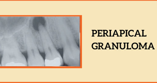
Periapical Granuloma, commonly known as Dental Granuloma, usually occurs at the tip of the root. When the infection is untreated for an extended period, it develops into a dental granuloma.
In the following blog, we’ll discuss the symptoms, causes, and management options for Periapical Granuloma, as well as preventive measures.
What is Periapical/Dental Granuloma?
When tooth decay is neglected for an extended period, the infection infects the tooth’s pulp. Eventually, it spreads to the periapical region, which surrounds the tooth’s root tip and contains bone, nerves, blood vessels, etc. This condition is known as periapical granuloma.
Although it can also affect males, it is more likely to impact females (>50%). Front teeth (incisors and canines) are more susceptible to this condition than teeth present at the back (premolars and molars). It can affect any tooth in either the upper or lower jaw. But often, the teeth of the upper jaw are affected.
Symptoms of Periapical Granuloma
- Tooth discoloration : The involved tooth is usually darkened and loses its natural color (white or yellow)—largely decayed tooth where food lodges in the tooth.
- Pain : Pain and discomfort can be experienced while eating or drinking food.
- Swelling : A swelling can be felt below the gums of the tooth, which can be present on the cheek or tongue side.
- Pus : Sometimes, pus discharges from the sinus (tract from the root tip towards the gums), which can also lead to a bad taste in the mouth.
How is periapical granuloma caused?
- Poor oral hygiene
- Decay of tooth
- Untreated cavity allowing infection/bacteria to reach the pulp and periapical tissues.
- Injury to the tooth
- Crack or fracture of the tooth surface, facilitating bacterial infiltration and inflammation in the periapical area.
Diagnosis of Dental Granuloma
- Dental caries are black or brown spots on the tooth surface.
- A dental x-ray (IOPA) is required to confirm this disease.
- Microscopic examination for significant swelling is necessary to diagnose periapical diseases.
Treatment Options for Periapical Granuloma
A simple dental filling can stop this process at an early stage. If a decayed tooth is left untreated, then root canal therapy is most probably required. If the swelling is large and the tooth cannot be saved, it has to be removed from the jaw (Dental tooth extraction).
Why is it important to treat it early?
Treating dental granulomas promptly is essential for several reasons, including:
- Timely treatment of dental granulomas prevents complications like abscesses and cysts.
- Early intervention relieves pain and discomfort for the patient.
- It preserves the affected tooth, avoiding the need for invasive procedures like root canals.
- Prevents the spread of infection to neighboring teeth or into the bloodstream.
- Better prognosis and faster resolution with early treatment.
- Cost-effective compared to managing advanced infections.
- Failure to treat promptly can lead to periapical cysts and jaw defects.
Preventive Measures for Periapical Granuloma
- Pit and fissure sealants prevent caries when applied by a dentist.
- Regular dental checkups aid in early caries detection.
- Dental fillings remove infection and halt decay.
- Root canal therapy protects periapical tissues if done promptly.
- Fluoride application on children’s teeth prevents decay.
What is the difference between a dental cyst and a dental granuloma?
| Feature | Dental Cyst | Dental Granuloma |
|---|---|---|
| Definition | Sac filled with fluid material/pus. It is located in the jaw bone. | Localized inflammation caused by chronic irritation or infection in the tooth root. |
| Location | Present in the jawbone near the tooth root. | Found at the tip of the tooth’s root or in the surrounding bone. |
| Origin | Arises from the epithelial remnants of the tooth’s developmental process or from the epithelial lining of the dental follicle. | Develops from chronic irritation or infection that triggers an immune response. |
| Appearance | Radiographically, appears as a round or oval | Radiographically, appears as a radiolucent |
| radiolucency with well-defined borders. | lesion with less-defined borders. | |
| Symptoms | May be asymptomatic or present with swelling, | May present with persistent pain, swelling, |
| pain, and occasionally drainage of pus. | and sometimes drainage of pus. | |
| Treatment | Usually requires surgical removal or | Treatment often involves endodontic therapy, |
| endodontic treatment if associated with a tooth | antibiotics, and sometimes surgical | |
| infection. | intervention if conservative measures fail. | |
| Prognosis | Typically good with appropriate treatment, | Prognosis depends on the extent of damage |
| but recurrence can occur if not completely | and response to treatment. In some cases, | |
| removed. | surgical intervention may be necessary. | |
| Common Causes | Infection due to tooth decay, trauma, or | Chronic dental infections, trauma, or |
| obstruction of salivary glands. | inflammatory processes affecting the tooth. |
Conclusion
Periapical granuloma can lead to tooth loss which can easily be saved by being vigilant about your oral health. Prevention can be done by means of fluoride application, pit and fissure sealants and regular dental check ups at Clove Dental. Dental fillings and root canal therapy at the earliest is essential to save the tooth.
References:
- Banomyong D, Arayasantiparb R, Sirakulwat K, Kasemsuwan J, Chirarom N, Laopan N, Lapthanasupkul P. Association between Clinical/Radiographic Characteristics and Histopathological Diagnoses of Periapical Granuloma and Cyst. Eur J Dent. 2023 Jan 4. doi: 10.1055/s-0042-1759489. Epub ahead of print. PMID: 36599448.
- Couto AMD, Meirelles DP, Valeriano AT, Almeida DS, Moraes Ê, Tarquinio SBC, Batista AC, MendonÇa EF, Costa NDL, Alves PM, Nonaka CFW, Abreu LG, Aguiar MCF. Chronic inflammatory periapical diseases: a Brazilian multicenter study of 10,381 cases and literature review. Braz Oral Res. 2021 Mar 15;35:e033. doi: 10.1590/1807-3107bor-2021.vol35.0033. PMID: 33729278.
- Moraes Ê, Tarquinio SBC, Batista AC, MendonÇa EF, Costa NDL, Alves PM, Nonaka CFW, Abreu LG, Aguiar MCF. Chronic inflammatory periapical diseases: a Brazilian multicenter study of 10,381 cases and literature review. Braz Oral Res. 2021 Mar 15;35:e033. doi: 10.1590/1807-3107bor-2021.vol35.0033. PMID: 33729278.
Dr. Dipanshu Aggarwal
Leave a Reply
Leave a Reply