


Dental Granuloma – Symptoms, Causes, Treatment
Medically Reviewed By Clove Dental Team
Written By
Dr. Nayanika Batra
Last Updated 27 April 2025
Periapical Granuloma, which is popularly known as Dental Granuloma, is a chronic inflammation that develops on the root apex. It is mostly asymptomatic in its early stages but can silently progress leading to discomfort and swelling causing pain.
In the following blog, we will be reading about the symptoms, causes, and options to manage Periapical Granuloma. We will also talk about the prevention measures and why it is important to treat it before it progresses.
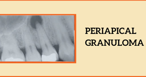
What is Periapical/Dental Granuloma?
Periapical granuloma, also referred to as a radicular granuloma, apical granuloma or simply dental granuloma, is an inflammation that occurs at the tip of an infected tooth.
It is a lesion or mass that typically starts out as an epithelial lined cyst, and undergoes an inward curvature that results in inflammation of granulation tissue at the root tips of a dead tooth.
Studies show that it is more common in females as compared to males. Anterior teeth (front teeth) are more predisposed compared to posterior teeth (back teeth). Periapical Granuloma can occur in any tooth but it commonly occurs in teeth of the upper jaw.
Symptoms of Periapical Granuloma
- Tooth discoloration: The concerned tooth is usually blackened, and it becomes discolored from its original color, white or yellow.
- Pain: Pain and discomfort can be felt at the time of eating or drinking
- Swelling: Swelling is present towards the side of the gum , which can be either on the cheek side or towards the tongue side. This may indicate the presence of an active infection.
- Pus: Sometimes, pus oozes out of the sinus, or tract from the root tip towards the gums. This indicates abscess formation in Granuloma.
How is periapical granuloma caused?
- Poor oral hygiene
- Decay of tooth
- Untreated cavity letting infection/bacteria enter to the pulp and periapical tissues
- Injury of the tooth
- Crack or fracture of the tooth surface, letting bacteria in to infect and cause inflammation in the periapical area
Diagnosis of Dental Granuloma
- Dental caries will look like black or brown spots on the surface of the tooth.
- The infection should be confirmed by a dental x-ray or IOPA.
Treatment Options for Periapical Granuloma
- Root Canal Treatment (RCT): Cleans and seals the root canal to remove infection.
- Apicoectomy: Surgically removes the root tip and infected tissue if RCT fails.
- Tooth Extraction: Done if the tooth cannot be saved, followed by replacement options.
- Medications: Antibiotics for severe infection; pain relievers for symptom control.
- Preventive Care: Maintain oral hygiene and address dental issues early.Success depends on timely treatment and proper care.
Why is it important to treat it early?
- Treatment of dental granulomas at an early stage prevents complications like abscesses and cysts.
- Early treatment reduces the amount of pain and discomfort to the patient.
- The lesion does not spread to adjacent teeth or the blood stream.
- Prognosis is good with early treatment, it resolves early.
- Cost effective before advanced infections develop.
- If not treated early then, it results in Periapical cyst and jaw defects.
Prevention from Periapical Granuloma
- Dental fillings remove infection, and the decay is arrested.
- Root canal therapy preserves the tissues in the periapical region if it is done in time.
- Fluoride Application in children to prevent cavity formation thus avoiding further complications.
Conclusion
Periapical granuloma, or dental granuloma, is a chronic inflammatory condition that occurs at the root tip of a dead tooth, often caused by untreated decay, trauma, or poor oral hygiene. Maintaining good oral hygiene, addressing cavities promptly, and regular dental check-ups can help prevent periapical granulomas. Early intervention ensures less invasive treatments, preserves oral health, and reduces overall costs and discomfort. Recognizing symptoms like tooth discoloration, pain, or swelling is essential to seek timely care and achieve favorable outcomes.
If you/someone you know has any symptoms related to Periapical Granuloma, make sure you visit your nearest Clove Dental Clinic.
Leave a Reply
Leave a Reply
Expert Guidance for Healthy Smiles
Book your free consultation today and take the first step towards healthy, beautiful teeth.
Membership Plans
Special thanks to our 30 Lakh valued customers
Dental Health
Plan Premium
Buy and Get Benefits at
590/-
in dental Treatments
Explore More Similar Posts
Explore More Blogs


Leave a Reply